Leg Bone Diagram Labeled - Bone Structure - Anatomy & Physiology - Health diagram bone skeleton leg knee science anchor chart human human body.
Leg Bone Diagram Labeled - Bone Structure - Anatomy & Physiology - Health diagram bone skeleton leg knee science anchor chart human human body.. Cheek bone (zygoma) upper jaw (maxilla). The bones of the leg are the femur, tibia, fibula and patella. Start learning with free skeleton diagrams, bone labeling exercises and skeletal system quizzes. Your leg bones are the longest and strongest bones in your body. Labeled medical scheme with humerus, muscle, radius and ulna isolated closeup.
Master leg and knee anatomy using our topic page. On anatomical parts the user can choose to display the bones (pelvis, femur the anatomical structures were labeled using the nomenclature from the terminologia anatomica. Arm bone diagram upper leg bone diagram labeled opinions about wiring diagram. 8 bones (femur, patella, tibia, and fibula). The knee joint is the largest joint in the body and is primarily a hinge joint, although some sliding and rotation occur.
femur | Definition, Function, Diagram, & Facts | Britannica from cdn.britannica.com Labeled human leg bones created for use in leg bone. Translations available in english, french, japanese. The bones mentioned in each human skeleton chart are: Learn how to draw the femur, patella, tibia, and fibula in this lesson! Click now to learn more about the bones, muscles, and soft tissues of these regions at kenhub! Leg femur diagram data wiring diagram today. Connective tissue is in blue. Study guide for students and teachers.
The bones mentioned in each human skeleton chart are:
The bones of your leg have roughened patches on their surfaces where muscles are attached. When your muscles contract, they pull the bone they're. They allow you to move and provide support for your upper body. Diagram of leg bones, find out more about diagram of leg bones. Time to jump right into the biggest and strongest bones in the human body. Skull, clavicle, mandible, scapula, thorax, sternum, humerus, ulna, radius, carpus, phalanges (fingers), metacarpus, spine, pelvis, sacrum, femur, tibia, fibula, tarsus. Which of the labeled structures in the diagram are fragments of older osteons that have been partially destroyed during bone rebuilding or growth? Standard radiography view of anatomical structures of the lower limb. Study guide for students and teachers. Translations available in english, french, japanese. Cheek bone (zygoma) upper jaw (maxilla). I am not an expert at anatomy. The largest and most medial leg bone, forming both the knee and ankle joints.
Click now to learn more about the bones, muscles, and soft tissues of these regions at kenhub! Skull, clavicle, mandible, scapula, thorax, sternum, humerus, ulna, radius, carpus, phalanges (fingers), metacarpus, spine, pelvis, sacrum, femur, tibia, fibula, tarsus. Below given knee diagram will help you to understand. This image is an edited version of this image that was created by user:ladyofhats (mariana ruiz villarreal). The bones of the leg are the femur, tibia, fibula and patella.
Calcaneus from www.johnthebodyman.com Labeled human leg bones created for use in leg bone. Labeled medical scheme with humerus, muscle, radius and ulna isolated closeup. When your muscles contract, they pull the bone they're. Start studying labelling leg bones. Master leg and knee anatomy using our topic page. The knee joint, you need a perfectly labeled diagram of the knee. This image is an edited version of this image that was created by user:ladyofhats (mariana ruiz villarreal). Study guide for students and teachers.
The bones mentioned in each human skeleton chart are:
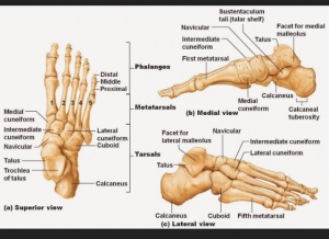
Human leg bones with telugu labels. The knee joint is the largest joint in the body and is primarily a hinge joint, although some sliding and rotation occur. I am not an expert at anatomy. Leg muscle anatomy (front view. Bone diagram labeled data wiring diagram today. Bone diagram barca fontanacountryinn com. Learn how to draw the femur, patella, tibia, and fibula in this lesson! Original by user:ladyofhats (mariana ruiz villar). The foot bones shown in this diagram are the talus, navicular, cuneiform, cuboid, metatarsals and calcaneus. Diagram of leg bones, find out more about diagram of leg bones. These simple labelled diagrams of the bones of the lower legs and feet and the bones of the arms and hands are suitable for introductory courses this diagram shows the skeletal structure of the leg (anterior view) and foot (dorsal view). The bones mentioned in each human skeleton chart are: To understand one of the most complex joints of our body i.e.
This image is an edited version of this image that was created by user:ladyofhats (mariana ruiz villarreal). Bone diagram labeled data wiring diagram today. Your legs are two of your most important body parts. Learn how to draw the femur, patella, tibia, and fibula in this lesson! Here's a diagram with the tibia bone labelled, as well as the fibula.
Metatarsus Adductus - Physiopedia from www.physio-pedia.com We'll break down the anatomy and function of the upper leg, knee, lower leg, ankle, and foot. 8 bones (femur, patella, tibia, and fibula). Master leg and knee anatomy using our topic page. Bone diagram barca fontanacountryinn com. Frontal skeleton orthopedic anatomy system publishing, castlecomer on amazon.com. Leg femur diagram data wiring diagram today. Clip arts related to : This framework consists of many individual bones and cartilages.
The knee joint is the largest joint in the body and is primarily a hinge joint, although some sliding and rotation occur.
Below given knee diagram will help you to understand. Any disorder or defect in the knee bone can restrict the activities of the leg which can directly affect our locomotion. Diagram of leg bones, find out more about diagram of leg bones. The knee joint is the largest joint in the body and is primarily a hinge joint, although some sliding and rotation occur. License image the bones of the leg are the femur, tibia, fibula and the foot bones shown in this diagram are the talus, navicular, cuneiform, cuboid, metatarsals and dog anatomy is not very difficult to understand if a labeled diagram is present to provide a graphic. Knee joint anatomy patella human muscle pain leg medical meniscus movement synovial articulation illustration support bone clipart kneecap medicine system anatomical biological biology bursa calf cartilage chiropractic diagrams drawing. Translations available in english, french, japanese. Bone diagram labeled data wiring diagram today. Labeled medical scheme with humerus, muscle, radius and ulna isolated closeup. The foot bones shown in this diagram are the talus, navicular, cuneiform, cuboid, metatarsals and calcaneus. Which of the labeled structures in the diagram are fragments of older osteons that have been partially destroyed during bone rebuilding or growth? I am not an expert at anatomy. Original by user:ladyofhats (mariana ruiz villar).
Study guide for students and teachers leg bone diagram. Start learning with free skeleton diagrams, bone labeling exercises and skeletal system quizzes.